Details
Start:
November 23, 2021
9:00 AM
(UTC/GMT +01:00 - Europe / Lisbon)
End: November 25, 2021
1:00 PM
(UTC/GMT +01:00 - Europe / Lisbon)
IGC
Speakers
Exciting lineup of speakers and community workshop presenters for the 2021 edition of SPAOM:
Paul French
Vice Dean (Research) for the Faculty of Natural Sciences at Imperial College London
Photonics Group, Physics Department, Imperial College London

Guillaume Jacquemet
PI of the Cell Migration Lab
Åbo Akademi University - Turku
Caren Norden
Group Leader of the Cell Biology of Tissue Morphogenesis Lab at IGC
Instituto Gulbenkian Ciência - Oeiras
Edgar Gomes
Group Leader
iMM - Institute of Molecular Medicine - Lisbon

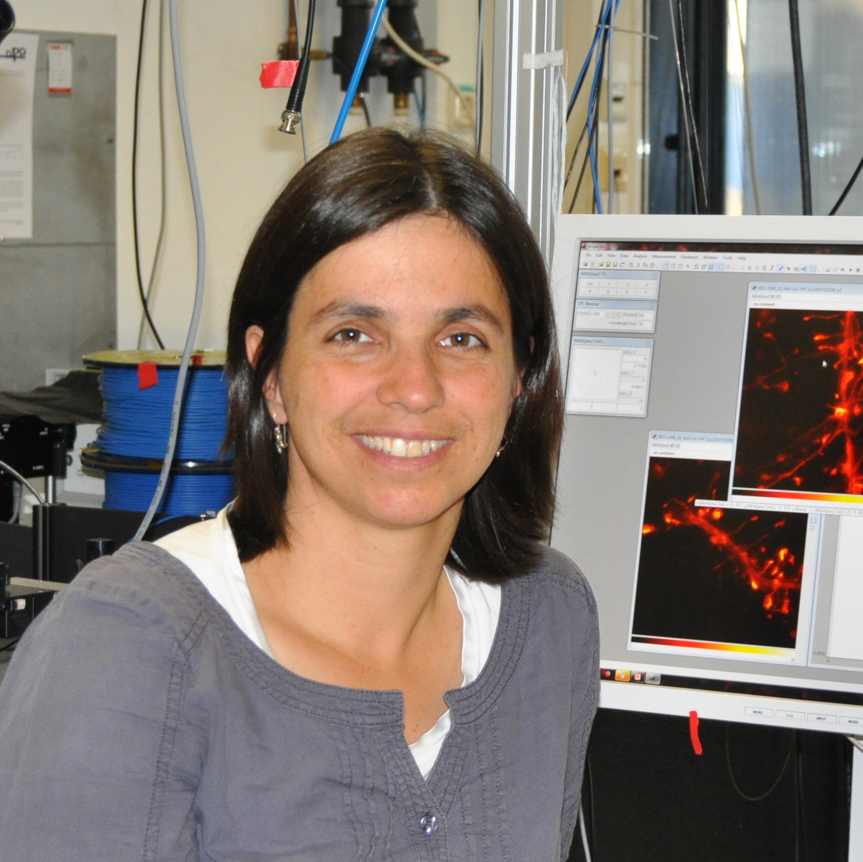
Katrin Willig
Group Leader
Max Planck Institut of Experimental Medicine, Göttingen
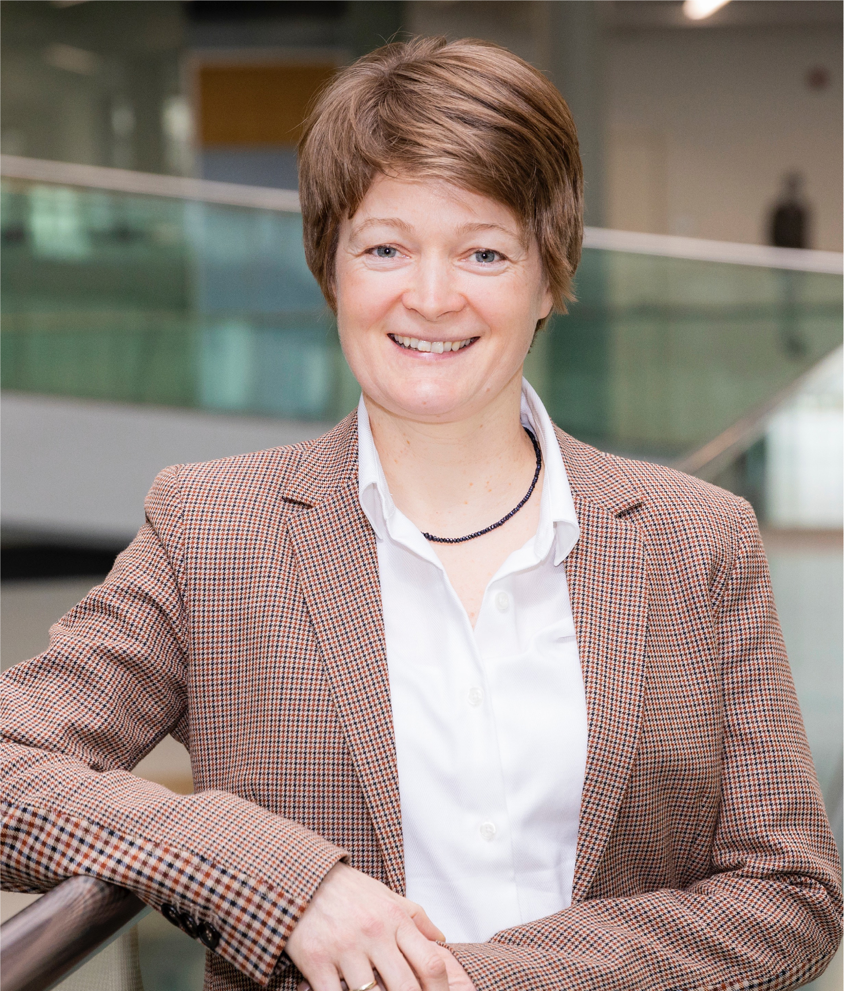
Vera Kozjak Pavlovic
Chair of Microbiology Biocenter
University of Würzburg


Erin Tranfield
Electron Microscopy Facility Head
Electron Microscopy Facility - Instituto Gulbenkian de Ciencia
Ana Laura Sousa
Electron Microscopy Facility Senior Technician
Electron Microscopy Facility - Instituto Gulbenkian de Ciencia
Community workshop guest speakers:


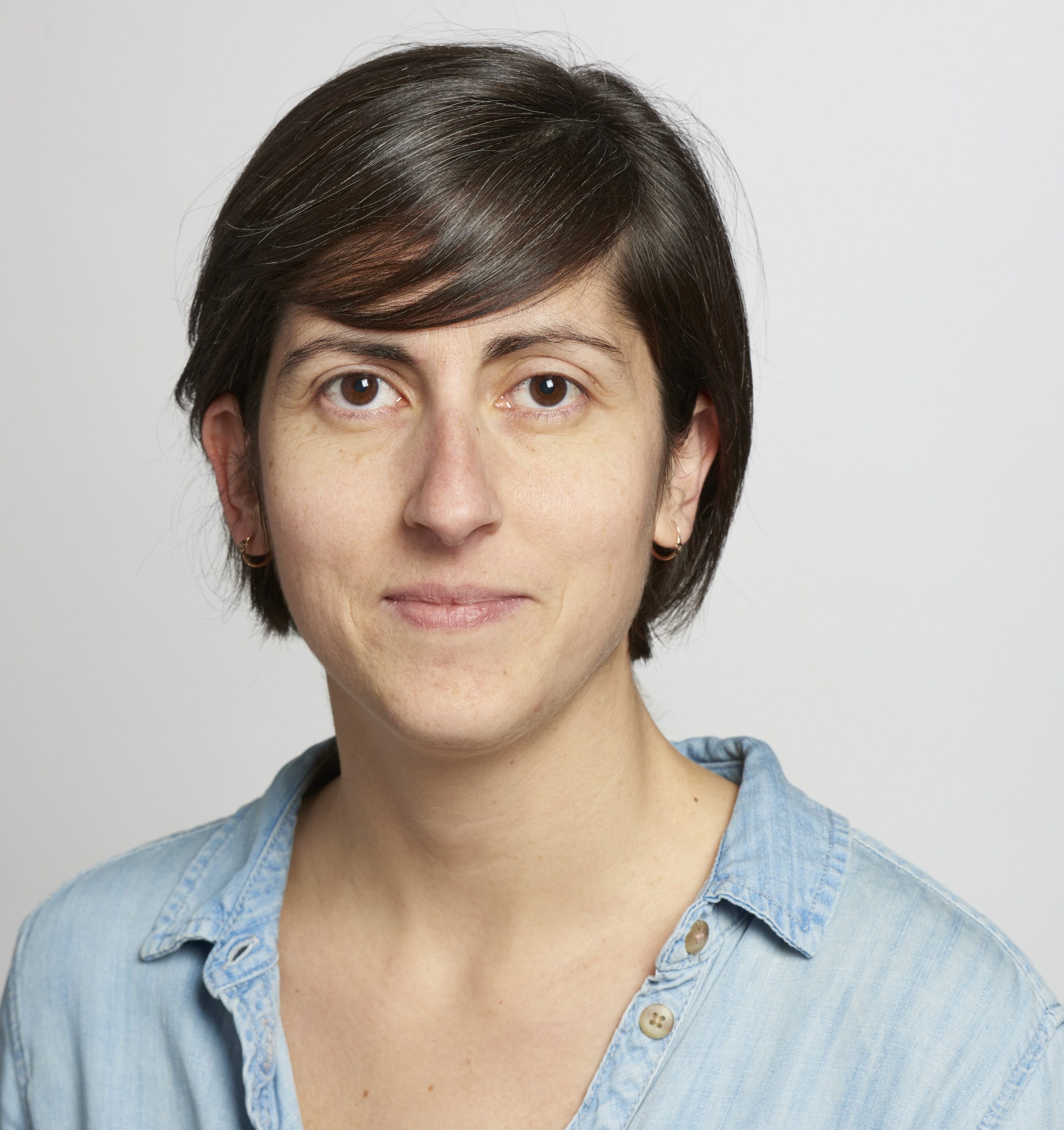
Anna Oddone
Community workshop - Frugal bioimaging
Universidad Pompeu Fabra, Barcelona, SPAIN


Marc Bickle
Community workshop - HTM/HCS
Max Planck Institute of Molecular Cell Biology and Genetics - Dresden

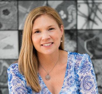
Claire Brown
Community workshop - Phototoxicity
McGill University - Advanced Bioimaging Facility
Johanna Bischof
Community Workshop - Remote Access
EuroBioimaging Bio-Hub | EMBL

Sponsors
The SPAOM2021 organizers gratefully acknowledge the support of our sponsors:


Organizers
Alexandre Lopes
OPenT/OpenSpin developer and Microscopy Assistant | UIC - Advanced Imaging Unit | Instituto Gulbenkian de Ciência
Gaby G. Martins
Facility Head | UIC - Advanced Imaging Unit | Instituto Gulbenkian de Ciência
Paula Sampaio
Advanced Light Microscopy Platform Head | Instituto de Investigação e Inovação em Saúde | Universidade do Porto
Scentific Committee
Hugo Botelho
Researcher | Faculty of Sciences of the University of Lisbon
José Rino
Head of the Bioimaging Unit | Instituto de Medicina Molecular João Lobo Antunes.
Maria Calvo
University of Barcelona

Ricardo Henriques
Group Leader at Instituto Gulbenkian de Ciência, Honorary Professor at University College London and Francis Crick Institute
Institutional Support & Logistics


PPBI support